Click here to return to newsletter contents.
Late in the autumn of 2001 I was sitting at my desk in the Museum of Anthropology drafting my schedule for the winter. I was nearly finished with all of my engineering prerequisites and was reading about what classes I was supposed to take next semester. To my horror, I realized that I would be spending nearly all of my time on North Campus, deprived of my LS&A world and my peers in the Ruthven Museum. I conveyed this misfortune to my boss, Karen O'Brien, and pointed out that if I spent all of my time on North Campus, I just might go crazy. I decided the only way to prevent such a fate was to devise a project involving both worlds. Thus was born the mummy project.
According to Karen, the Museum of Anthropology was in possession of a mummy child. At that point the details regarding its condition and location were unclear, and to my dismay I learned several weeks later that the child had already been unwrapped.
But the idea was stuck in the back of my head, and after another week I started pursuing other options. At that point, I recalled that my former classical archaeology professor, Dr. Susan Alcock, worked as a Research Scientist in the Kelsey Museum of Archaeology. The very same day I called her, and though admitting it was a little far fetched, she agreed to approach the board of curators with my proposal that month. By chance, I also met the Acting Director of the Kelsey, Dr. Lauren Talalay, and pitched the idea to her. She verified that the Kelsey had a mummy child and, together with Dr. Alcock, agreed to approach the curators.
In the meantime, I had to find someone who could take CT scans for me and an advisor in the Materials Science and Engineering (MSE) Department. Karen had mentioned that she knew someone in Radiology who could take x-rays for me, so I began by wandering down to Radiology. Four hours later, after talking to a dozen hospital workers, walking through six buildings, and visiting a Wendy's, I ended up on the opposite corner of the Medical Campus in the Computer-aided Tomography (CT) Department at the mercy of a kind old secretary, Rosita. She introduced me to the Associate Chair of Research, Dr. Isaac Francis, and the head technician, Kim Pici. Over the next four weeks I would become intimately acquainted with them and would eventually coax them into letting me bring the mummy in after hours to get scans. In these four weeks I had also managed to find a mentor in the MSE Department, Dr. John Halloran, from whom came an infinite well of advice and support.
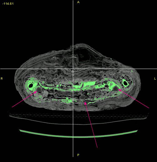
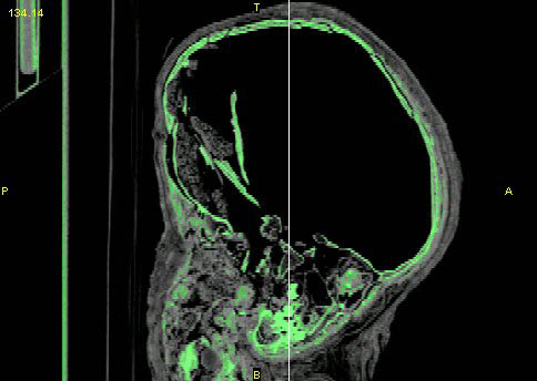
Permission from the Kelsey was granted in late January. Two Curators of Ancient Egypt from the Kelsey, Dr. Terry Wilfong and Dr. Janet Richards, volunteered to accompany me in to the hospital, and on February 19 the triumvirate of two curators and one student brought the Kelsey Museum's mummy to the University of Michigan hospital and completed one axial CT scan. Without a scratch, the mummy returned to its home on the third floor of the Kelsey later that night.
Now that I have the data, it is my responsibility to interpret it. I was fortunate enough to obtain powerful medical imaging software with a limited license and, with it, have started to build three-dimensional computer models. It is a surprisingly detailed process, and over the course of two weeks I have only been able to separate the legs and arms. Hopefully, within the bounds of my temporary software license I will be able to build an accurate representation of the skull and as much of the body as possible. These data sets will be saved in a format known as stereolithography (STL) data.
|
|
From these data, I will attempt to build accurate plastic representations of my computer models in what is known as a stereolithographic apparatus, or SLA. Under the guidance of Dr. Halloran, a model accurate to 13 nanometers should be producible. This model should provide scientists an accurate view of the child's bones. From it, we will be able to study diet, age, and countless other aspects of the child's life--all without ever touching the mummy.
I have been blessed with a very patient and forgiving group of scientists to help me. I would like to extend my deepest thanks to John Halloran, my advisor; Karen O'Brien, Collections Manager at the Museum of Anthropology; Terry Wilfong and Janet Richards, Curators of Ancient Egypt at the Kelsey Museum; Lauren Talalay, Acting Director of the Kelsey; Susan Alcock, Research Scientist at the Kelsey; Isaac Francis, Associate Chair of Research and friend in CT; Kim Pici and Karen Overbay, CT technicians; and Bill Marlette and Tom Hulscher at GE. Your benevolence has made this project what it is.
Grant K. M. Martin, Undergraduate
School of Engineering
Copyright © 2002 The Kelsey Museum of Archaeology, University of Michigan. All rights reserved.