
Click here to return to newsletter contents.
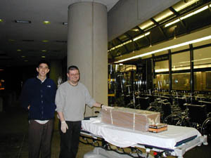
On the cold, dark night of February 19, we met at the Kelsey Museum for a most unusual errand. We were about to transport a body to the hospital.
 |
Of course, this body was one of our two human mummies (Kelsey Museum inv. 1971.02.0179), the mummy of a boy from the Roman period, probably just under 2,000 years old. This mummy was donated in the late 1800s to the Bay View Association, in Bay View, Michigan, by a Miss Hattie M. Conner, of Cairo, Egypt. In 1971, the Bay View Collection was acquired by the Kelsey Museum, and the mummy has been in Ann Arbor ever since. He was last on view in 1995 in the turret gallery, as part of the exhibition "Preserving Eternity," but ultimately his condition became a cause for concern and he was taken off display. He now resides in a special cabinet in our climate-controlled storage area. Thanks to the initiative of U-M Engineering undergraduate Grant Martin, we were taking this mummy to the hospital for a series of CT scans--as a way of looking inside the mummy without damaging it.
Grant had proposed to use CT technology to create a 3-D model of the child mummy, the data from which could be transferred into a CAD program and organized by density and layer so that we could have a series of "views" of the mummy. The mummy had already been x-rayed by our local mummy expert James Harris back in the 1970s; the x-rays show the mummy to have been a boy of probably 10 to 12 years but don't provide much more information. We were hoping that the CT scans would allow us to learn much more about this Egyptian boy of the Roman period.
Preparation for this trip involved a number of Kelsey Museum people. A big thank-you to our conservator, Suzanne Davis, who in the midst of her incredibly busy time working on the Cavafy exhibition managed to prepare the mummy for his visit to the hospital, and thanks to IPCAA graduate student Molly Swetnam-Burland for helping Suzanne. Thanks also to Kelsey Museum Registrar Robin Meador-Woodruff for taking care of the paperwork; Robin's careful attention to the necessary details involved in transporting a mummy around town made us feel a little more confident that we could manage to stay out of jail if stopped for carting around what was, after all, a dead person.
Grant picked us and the mummy up at 10 p.m. in a minivan borrowed from one of the local funeral homes (!); we rode in the back with the mummy, trying not to think of all the other passengers who had taken a similar ride in this vehicle, while Grant drove us to the hospital. During the day, the University of Michigan Hospital has many entrances and means of access; by the time we arrived, most of the doors were locked, so we got to spend some quality time with the mummy (safely packed in a specially designed box and wrapped in plastic) while Grant frantically searched for a way to get in. Once inside, we made the surprising discovery that many of the hospital elevators would not accommodate the gurney we had commandeered. Eventually, we found our way to the Radiology Department.
 |
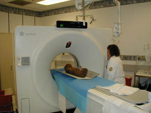
Karen Overbay, a CT technician, met us there, and we rapidly collected a crowd of late-shift doctors, nurses, engineers, and other technicians ("I heard you were CT-ing a MUMMY in here!!"). (As you can see, we captured all the excitement on the Abydos digital camera, competing for photo ops with Grant's camera). Benefiting from these high levels of local interest, the child mummy ended up traveling through the best CT scanner in the hospital. As the mummy, looking rather small and forlorn amidst all the technology, passed back and forth through the equipment, ghostly images appeared and changed on the technician's monitor. The results that we could see on screen were phenomenal.
 |
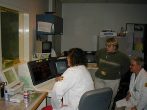 |
The child was clearly very expensively and carefully treated; innumerable layers of fine linen were visible in the CT sections, and the evidence indicates that his organs and brain were removed (fragments of bone could be seen inside the skull cavity, probably driven there by the embalming probe used to extract the brain). The results of this scan and modeling should also provide a wealth of information on his health status and dietary habits--the level of detail visible on the teeth, for example, was amazing. The positioning of the body within its wrappings was interesting to see: the boy's head was tilted down, and the body seems to have been compressed under the weight or pressure of the many layers of bandages.
Perhaps the most startling fact to emerge from watching the scans as they were happening was that the child seems to have six fingers on one of his hands. (You can imagine the furor in the viewing/computer room when this discovery was made.) Excessive fingers or toes (polydactyly) is an unusual condition in humans that can be caused by a number of factors. In Roman Egypt, however, cases of polydactyly may well have been a genetic consequence of the high incidence of brother-sister marriage among the population at large--one of the few populations in human history in which this traditionally taboo practice was widespread. (Thanks to Walter Scheidel at the University of Chicago for his insight into these matters.) Although DNA testing of mummies has garnered mixed results, it might be that such tests on our mummy could confirm or disprove this hypothesis.
After several passes through the machine, the mummy was fully scanned. He seemed to have suffered no ill effects from the procedure, and we carefully packed him up and headed back to the Kelsey Museum. By a stroke of good luck, we arrived at the Museum just in time to get the mummy back to his resting place in the SAFE before the entire building master-armed itself. The mummy survived the trip in good shape, and Grant Martin is now working on the data to create his 3-D models (see the report from Grant on page 8). We are thinking about ways to make the information ultimately gained from this project available to our visitors, both in the gallery and on our website. It's a tricky balance, satisfying our scientific curiosity while trying to maintain respect for the wishes of the ancient Egyptians, but we think that noninvasive means of investigation like CT scanning provide the best compromise. Whatever happens, we'll keep you posted!
Janet Richards and Terry Wilfong
Copyright © 2002 The Kelsey Museum of Archaeology, University of Michigan. All rights reserved.